
I bet you’ve said “I need to get this mole checked out sometime.” About once a week, a patient will show me an ugly looking mole that gives me no concern for cancer (I’ll give you the 411 on these specific buggers in a bit). Occasionally, a patient will say, “does this look concerning to you?” and I truly don’t know what to tell them. I want to say, “eh it looks fine, keep an eye on it…” but that wouldn’t always be responsible of me. PCPs know the general appearance of (pre)cancerous moles, but some skin cancers are sneaky, so I refer these patients to see a dermatologist for an opinion/ biopsy. I never take a chance on a mole that worries a patient. Their peace of mind is worth the speciality visit. I remember one time I was listening to a patient’s heart with my stethoscope and I saw a tiny blue dot under their collarbone (size of a pen mark). They hadn’t thought anything of the mark, but I immediately placed a referral for dermatology — it turned out to be melanoma. Do primary care clinicians provide a head-to-toe skin exam at each and every office visit? No, probably not. But we do glance at the sun-exposed areas during other parts of the exam and will voice if we’re concerned about a mark or two. OK so how do you know that mole you’ve had for 25 years is harmless? How do you know that spot on your cheek could be worrisome? Let’s delve…
The most common skin spots (excluding rashes) that I see are the following, in order: Seborrheic keratoses (that ugly guy I mentioned earlier), actinic keratoses, and normal nevi/ skin tags (the technical term for mole is nevus). I’ll break each of these down, with photos! Yay!
Seborrheic keratoses are in my opinion one of the most unsightly skin lesions in existence. No, I’m not exaggerating. No offense if you have one, but most patients agree with me that they hate how this lesion looks and they want it GONE. Not only are they ugly, they can get caught on clothing, you can irritate one when drying off after your shower, or just graze it on something. They can bleed and hurt, which understandably alarms people for cancerous etiology. Rest assured— cancer arising from these growths isn’t something to worry about. DO NOT try to remove these on your own. They have a vascular supply and need to be handled in a Dermatology office setting. Some PCPs may do this, but if you have one “seb k” as I call them, you’ll likely have more. They tend to have a genetic pattern. I’ve had patients have at least 20 on their back at once. They’re slow growing, and get annoying over time. Does your dad have these on his back? You probably will, too, around 50+ years old. Get excited.
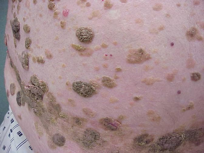
Source: MedScape
Actinic keratoses are lesions I considered placing in the “cancer” category later in the post, but that may be dramatic of me. Most squamous cell carcinomas arise from a lesion that was an actinic keratosis first, but not all AKs develop into cancer. So let’s talk about them here. They’re flaky, red/pink, rarely raised spots that tend to bleed a bit. They’re on sun-exposed areas. I see a lot of these on faces, scalps of balding men, and lower arms. These are normally seen on fair-skinned people. About 55% of white males 65+ have an actinic keratosis. Keep in mind that this age group didn’t utilize sunscreen in their younger years, so this places them at a heightened risk of skin damage. Treatment of these spots is normally topical with a cream you cannot get over-the-counter–it must be prescribed. If you have a crusty spot on your skin that gets itchy, flakes, bleeds a bit, sort of heals again, then irritates you a few weeks later, it could definitely be an AK. Get it looked at next time you visit your PCP or dermatologist.
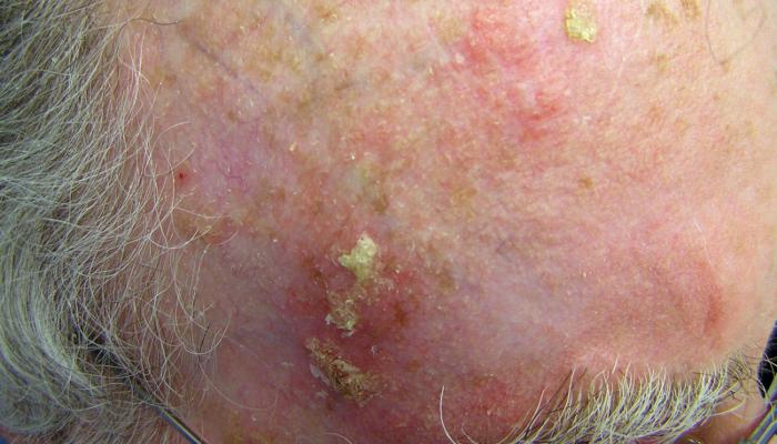
Source: https://contourderm.com/actinic-keratosis/

Source: https://www.webpathology.com/image.asp?case=703&n=18
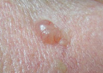
Benign nevi or the run-of-the-mill mole are super common and come in many shapes and sizes. A poorly placed mole can affect your appearance, and a beauty mark can help make your career (I’m looking at you, Cindy Crawford). As far as a non-concerning size, they’re normally less than 6 mm across. They can sometimes be flesh-colored, brown, or pinkish; flat or raised. There are several kinds of benign moles, so if you are concerned, just ask. Removing a mole because you’re worried it’ll be cancer one day isn’t necessary. Now, if it’s because you hate the look of it, a dermatologist or plastic surgeon may nicely do this for you, but insurance will laugh when you ask them to cover the procedure. Be prepared to pay out of pocket. (See the “Melanoma” section of this post to view a diagram/photos of benign, normal moles.)
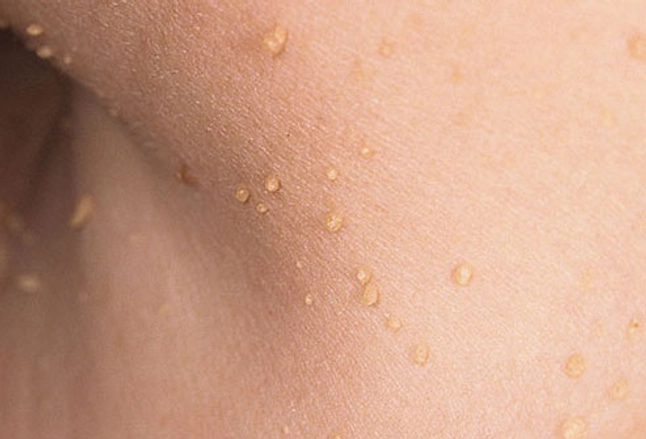
Skin tags occur mainly where lots of skin friction occurs. That may be your neck due to necklaces and shirt collars, under your arms due to arm/side of chest contact, and between the legs/buttocks for obvious reasons. Some women find they get them at the bra line, also, and under their breasts. People mainly bring them up in an appointment because they hate the look of them and they want them removed. Removal is almost always very simple, and only rarely requires a suture. The size of the skin tag’s base will determine if a suture is needed. This is another procedure you’ll need to prepare paying out of pocket for. It would behoove you to have several to remove at once rather than just one per visit, as the former will be more cost-efficient than the latter.
The skin is one of the most important lines of defense the body has. Remember, the skin is an organ, and has significant potential for damage and disease like the rest of our bodies. I rarely see skin cancers, but I do want to say skin cancer is more common than you think! Here are the skin cancers in order of prevalence:
Basal Cell Carcinoma– The most common type of skin cancer is BCC. Usually, the growth limits itself to the skin, but it’s known to go to the bone underneath. Men have a 30% increased risk compared to women, and those with white skin are at most risk. Studies show that your history of childhood sun exposure puts you at more risk for BCC over adult sun exposure. Getting a severe sunburn in your lifetime places you at twice the risk as someone who hasn’t been burnt badly. As far as tanning beds go, 650 folks with BCC and a tanning bed use history were studied— they had a 60% higher risk to develop their cancer compared to those who’d never tanned. Already have a BCC? You have a 50% chance of having another one within 5 years. Keep up with your regular dermatology appointments and speak up if you feel that one is worrying you!
Source: https://www.spotscreen.com.au/info-centre/skin-cancer-information/skin-cancer-types/basal-cell-carcinoma-bcc/
Squamous Cell Carcinoma– about 20-50% of skin cancers are SCC. Non-hispanic whites have the highest incidence of this type. You see these lesions on sun-exposed areas, obviously, and near chronic wounds or around scars (wound skin cells divide a lot and have been damaged, so this is why a cancer is easily found in this type of environment). UVB ray exposure is considered to be the main cause of this cancer, and if your occupation places you in the sun on a regular basis (construction, landscaping, lifeguard, etc), you have a two-fold risk increase. UVA rays also assist in the risk— a study that looked at psoriasis patients receiving UVA light therapy noted a 35-fold increase in SCC. Bottom line, both types of UV rays cause damage to skin cell DNA. If the DNA doesn’t get fixed by your body’s repair mechanisms, the skin cells divide uncontrollably, which is cancer.
:max_bytes(150000):strip_icc()/bd1-4257053ebaae4346b42ef07c0ad7a5c1.JPG)
Source: https://www.verywellhealth.com/squamous-cell-carcinoma-1068874
Melanoma– while being the least common skin cancer, it’s definitely considered the most serious and aggressive. Unfortunately, we all likely know someone who’s had melanoma— it can become stage 4 if not caught in time and spreads to other organs. What’s scary about this type is, most melanomas arise on the skin without an associated mole. In fact, only 29% of melanomas arise as a mole. A change in your ABCDEs is a great guide for melanoma concern. Is the lesion asymmetrical? Is the border changing/jagged? Is the color looking different? Has the size/diameter grown (more than 6 mm total size)? And E is “ugly duckling” and “Evolution.” Has the lesion evolved suddenly overall? Does this lesion look nothing like your other moles? Usually people have a gut feeling about a mole when it just doesn’t look right. Bring it up soon to your specialist. Also, if you can’t get an office visit for a few weeks, take pictures of the lesion. Pictures may catch some evolving characteristics and may help with diagnosis.

Source: https://www.spectrumdermatologyseattle.com/importance-early-detection-melanoma/
I’m writing this post during the COVID-19 quarantine, so after you read this, do a self-assessment of your skin. You probably have some time on your hands. You may need a partner to look closely at your back. Have good lighting, and if you see something questionable, ask your PCP or dermatologist to take a look. Based on the definitions and descriptions above, you’ll know if it’s emergent or worth a casual mention later.
I hope you feel more educated on benign versus concerning skin lesions. Now, make good choices and wear your sunscreen.
References:
Hunt R et al. Acquired melanocytic nevi (moles). 2020. Retrieved from https://www.uptodate.com/contents/acquired-melanocytic-nevi-moles?search=seborrheic%20keratosis&topicRef=5573&source=see_link.
Lim G and Asgari M. Epidemiology and risk factors for cutaneous squamous cell carcinoma. 2020. Retrieved from https://www.uptodate.com/contents/epidemiology-and-risk-factors-for-cutaneous-squamous-cell-carcinoma?search=skin%20cancer&source=search_result&selectedTitle=5~150&usage_type=default&display_rank=5.
Padilla RS. Epidemiology, natural history, and diagnosis of actinic keratosis. 2020. Retrieved from https://www.uptodate.com/contents/epidemiology-natural-history-and-diagnosis-of-actinic-keratosis?search=actinic%20keratosis&topicRef=5336&source=see_link.
Swetter S and Geller A. Melanoma: Clinical features and diagnosis. 2020. Retrieved from https://www.uptodate.com/contents/melanoma-clinical-features-and-diagnosis?search=melanoma&source=search_result&selectedTitle=1~150&usage_type=default&display_rank=1.
Wu P. Epidemiology, pathogenesis, and clinical features of basal cell carcinoma. 2020. Retrieved from https://www.uptodate.com/contents/epidemiology-pathogenesis-and-clinical-features-of-basal-cell-carcinoma?search=basal%20cell%20carcinoma&source=search_result&selectedTitle=1~150&usage_type=default&display_rank=1
Featured Image: https://www.getldi.com/how-to-detect-skin-cancer/
